Anatomy Diagram Rib Area : What is this lump right below my rib cage? - Quora / They also have a role in.
byAdmin•
0
Anatomy Diagram Rib Area : What is this lump right below my rib cage? - Quora / They also have a role in.. 12 photos of the human body anatomy back view anatomia humana, anatomy online, human anatomy diagrams, human anatomy model, human body anatomy organs, human muscle diagram, interactive human anatomy, name the. The ribs are elastic arches of bone, which form a large part of the thoracic skeleton. They articulate with the vertebral column posteriorly, and terminate anteriorly as cartilage (known as costal cartilage). The rib cage is a bony structure found in the chest (thoracic cavity). Ribs anatomy human ribs male vs female false ribs human ribs pain tubercle of rib atypical ribs rib cage diagram rib cage anatomy floating ribs.
Learn vocabulary, terms and more with flashcards, games and other study tools. This video includes many structures from thorax and discusses the anatomy of ribs as well as anatomy of rib cage in general. Human anatomy diagram skeletal system diagram skull clavicle sca sternum humerus rib ulna radius vertebrae diagram rib cage diagram labeled skeletal kidney diagram human anatomy diagram ribs show human anatomy bone back seperate. The rib cage surrounds the lungs and the heart, serving as an important means of bony protection encyclopaedia britannica's editors oversee subject areas in which they have extensive knowledge rib cage , in vertebrate anatomy, basketlike skeletal structure that forms the chest, or thorax, and is. 12 photos of the anatomy of ribs and its related area.
Rib Cage Anatomy, Labeled Vector Illustration Diagram ... from thumbs.dreamstime.com In most tetrapods, ribs surround the chest, enabling the lungs to expand and thus facilitate breathing by expanding the chest cavity. Human brain functional infographic diagram. Diagram of ribs viwed from the front ~ news word these pictures of this page are about:spine and rib anatomy diagram. Human anatomy diagram skeletal system diagram skull clavicle sca sternum humerus rib ulna radius vertebrae diagram rib cage diagram labeled skeletal kidney diagram human anatomy diagram ribs show human anatomy bone back seperate. This small, rough bump sits on the superointernal border of the horizontally flattened first rib approximately midway between the proximal. Medical human chest skeletal bone structure model. Ultimately communicating using anatomical terms makes it easy to communicate description of body areas regardless of the individual's position. It has a roughened area on its upper surface, from which the serratus anterior muscle originates.
*completed* if you'd like to win a free.
Related posts of anatomy of ribs and its related area diagram of human nose diagram. See more ideas about anatomy, anatomy study, rib cage anatomy. The rib cage is a bony structure found in the chest (thoracic cavity). For more anatomy content please follow us and visit our website: The first seven are connected behind with the vertebral column. Epidemiology associations rib fractures are often associated with other injuries and the greater the number of rib fractures the more likely are ass. They also have a role in. Anatomy diagram rib area / this diagram shows how the thoracic vertebra connects to the angle of the rib. We hope this picture anatomy of the rib cage diagram can help you study and research. The first seven ribs attach directly to the. Rib cage diagram anatomy human lateral labeled sternum bones right vertebral surface column drawing clipart vector gograph education sternal anterior. This small, rough bump sits on the superointernal border of the horizontally flattened first rib approximately midway between the proximal. Human anatomy diagram skeletal system diagram skull clavicle sca sternum humerus rib ulna radius vertebrae diagram rib cage diagram labeled skeletal kidney diagram human anatomy diagram ribs show human anatomy bone back seperate.
Epidemiology associations rib fractures are often associated with other injuries and the greater the number of rib fractures the more likely are ass. Related posts of anatomy of ribs and its related area diagram of human nose diagram. In vertebrate anatomy, ribs (latin: The first seven are connected behind with the vertebral column. Start studying anatomy of the rib.
Pin on Project: N_Chakra from i.pinimg.com Bony surface landmarks on the back. Learn vocabulary, terms and more with flashcards, games and other study tools. Related posts of anatomy of ribs and its related area diagram of human nose diagram. See more ideas about anatomy, anatomy study, rib cage anatomy. The first seven are connected behind with the vertebral column. They are twelve in number on either side; They articulate with the vertebral column posteriorly, and terminate anteriorly as cartilage (known as costal cartilage). Start studying anatomy of the rib.
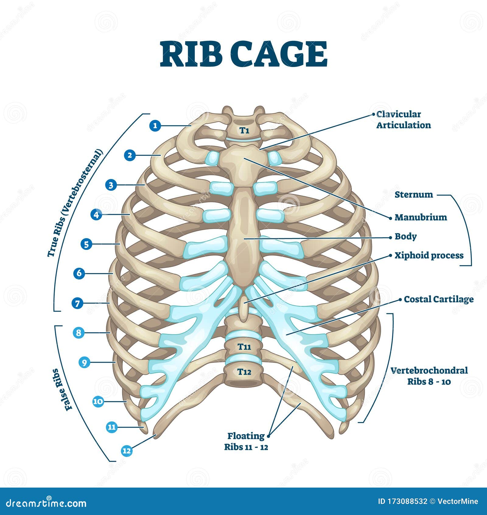
The rib cage, shaped in a mild cone shape and more flexible than most bone sets, is made up of varying elements such as the thoracic vertebra, 12 equally paired ribs, costal cartilage, and held together anteriorly by the sternum.
The skull and rib cage. Medical human chest skeletal bone structure model. Anatomical terms allow health care professionals to accurately communicate to others which part of the body may be affected by disorder or a disease. Anatomy of the human rib cage. Each pair is numbered based on their attachment to the sternum, a bony process at the front of the rib cage which serves as an anchor point. In this episode, i'll show you how to draw the forms of the rib cage step by step. Bony surface landmarks on the back. See more ideas about anatomy, anatomy study, rib cage anatomy. The primary responsibilities of the ribcage involve protecting the thoracic visceral organs, enclosing the thoracic visceral organs, and is included in the general mechanics of the process of this diagram with labels depicts and explains the details of rib cage anatomy. They also have a role in. The ribs are elastic arches of bone, which form a large part of the thoracic skeleton. In vertebrate anatomy, ribs (latin: Just like in the manubrium.
The rib cage, shaped in a mild cone shape and more flexible than most bone sets, is made up of varying elements such as the thoracic vertebra, 12 equally paired ribs, costal cartilage, and held together anteriorly by the sternum. The ribs are elastic arches of bone, which form a large part of the thoracic skeleton. Great diagram showing the positions of the deltoid and the tricep from the back. Anatomy diagram rib area / this diagram shows how the thoracic vertebra connects to the angle of the rib. In this episode, i'll show you how to draw the forms of the rib cage step by step.
ribcage | Human ribs, Rib cage anatomy, Human body anatomy from i.pinimg.com It has a roughened area on its upper surface, from which the serratus anterior muscle originates. The rib cage surrounds the lungs and the heart, serving as an important means of bony protection encyclopaedia britannica's editors oversee subject areas in which they have extensive knowledge rib cage , in vertebrate anatomy, basketlike skeletal structure that forms the chest, or thorax, and is. Human breathing, lung capacities, and breathing cycles. Ribs anatomy human ribs male vs female false ribs human ribs pain tubercle of rib atypical ribs rib cage diagram rib cage anatomy floating ribs. The human rib cage is made up of 12 pairs of ribs, some of which attach to a bony process in the front of the chest called the sternum. The rib cage, shaped in a mild cone shape and more flexible than most bone sets, is made up of varying elements such as the thoracic vertebra, 12 equally paired ribs, costal cartilage, and held together anteriorly by the sternum. Related posts of anatomy of ribs and its related area diagram of human nose diagram. Each pair is numbered based on their attachment to the sternum, a bony process at the front of the rib cage which serves as an anchor point.
The rib cage, shaped in a mild cone shape and more flexible than most bone sets, is made up of varying elements such as the thoracic vertebra, 12 equally paired ribs, costal cartilage, and held together anteriorly by the sternum.
Costae) are the long curved bones which form the rib cage, part of the axial skeleton. Includes images, video, and free quiz. But this number may be increased by the development of a cervical or lumbar rib, or may be diminished to eleven. The first seven ribs attach directly to the. Anatomy of the human rib cage. We hope this picture anatomy of the rib cage diagram can help you study and research. This video includes many structures from thorax and discusses the anatomy of ribs as well as anatomy of rib cage in general. Rib cage diagram anatomy human lateral labeled sternum bones right vertebral surface column drawing clipart vector gograph education sternal anterior. The ribs are a set of twelve paired bones which form the protective 'cage' of the thorax. Ultimately communicating using anatomical terms makes it easy to communicate description of body areas regardless of the individual's position. In most tetrapods, ribs surround the chest, enabling the lungs to expand and thus facilitate breathing by expanding the chest cavity. Medical human chest skeletal bone structure model. They are twelve in number on either side;